
Reading Time:
Overview
Flexion and extension occur at this hinge joint, in addition gliding, rolling and rotation are possible.
The knee is a common site of pathology and knee arthroplasty is the second most common joint replacement, the hip joint takes first spot.
Gross Anatomy
Articulations

Popliteus muscle
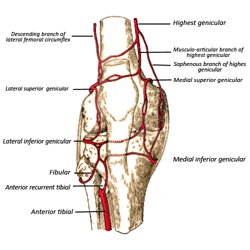
Blood supply
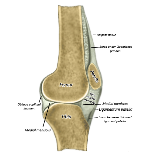
Suprapatella bursa - extends a hands breadth above the patellaPrepatella bursa - anterior to the patella, common site of irritationInfrapatella bursa - behind and adjacent to the patella tendon
The medial and lateral femoral condyles sit on their respective tibial condyles like two golf balls on adjacent golf tees. In order to deepen the articular surface of the tibia and help stabilise the joint, two fibrocartilaginous discs sit on top of the tibial plateau. Several "static" and "dynamic" stabilisers provide further support.
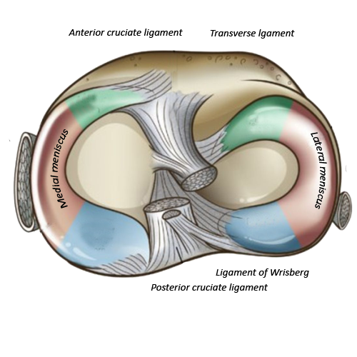
The menisci are wedge shaped pads that are attached to the margins of the tibial condyle by the coronary ligaments. The medial meniscus is C-shaped, its anterior horn attaches to the anterior intercondylar area, and the posterior horn attaches to the posterior intercondylar area.
The lateral meniscus is smaller and O-shaped and attaches to the intercondylar area. The anterior horns are attached by the transverse genicular ligament.
The patello-femoral joint is an articulation between the patella and the medial and lateral femoral condyles. The patella is a sesamoid bone and sits within the tendon of the quadriceps muscle. The quadriceps tendon inserts along the superior border of the patella and the patella ligament originates on the apex and inferior margins of the patella and inserts on the tibial tuberosity.
Joint capsule and extracapsular ligaments
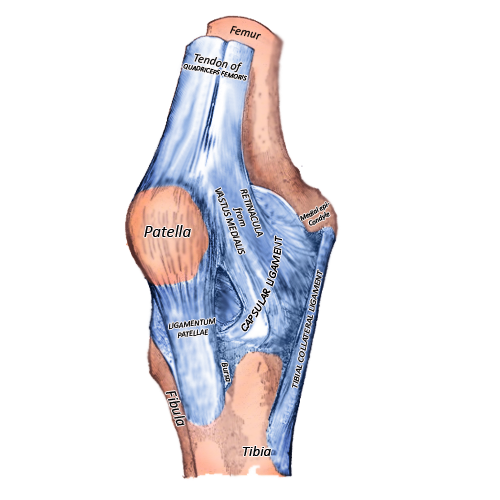
The joint is enveloped by a fibrous capsule that is incomplete in certain areas and thickened in others. These thickened areas form intrinsic ligaments which contribute to the stability of the joint. Superiorly it is attached to the femur, posteriorly to the femoral condyles and intercondylar fossa, and inferiorly to the tibial plateau. The tendon of popliteus passes through an opening in the posterior capsule. Anteriorly the capsule is replaced by the patella and quadriceps tendon and the adjacent retinacular bands. A synovial membrane lines the internal surface of the fibrous capsule.
"Extracapsular" ligaments include:
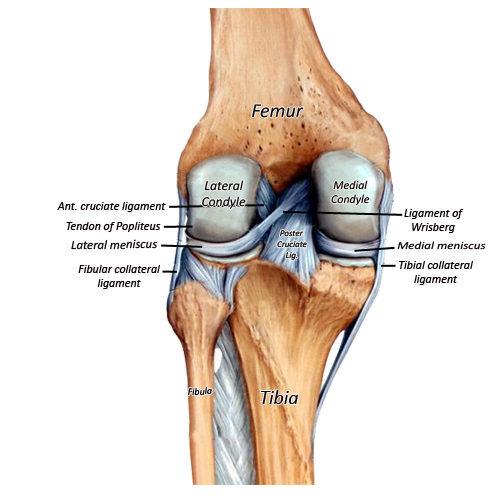
Fibular (lateral) collateral ligament - cord like structure from the lateral epicondyle to the fibular head
Tibial (medial) collateral ligament - band, femoral epicondyle and condyle to the superomedial tibia
Patella ligament - apex and inferior margins of the patella to the tibial tuberosity
Oblique popliteal ligament - posterior to the medial tibial condyle to the central part posterior capsule
Arcuate ligament - posterior fibular head to the posterior capsule
Cruciate ligaments
The cruciate ligaments, as demonstrated in the first image, cross over each other in an X formation.
The anterior cruciate ligament prevents anterior displacement of the tibia on the femur. It originates from the anterior intercondylar area and extends superiorly, posteriorly and laterally before inserting on the posterior part of the medial side of the lateral condyle of the femur.
The stronger posterior cruciate ligament prevents posterior displacement of the tibia on the femur. It originates from the posterior intercondylar area and inserts on the lateral surface of the medial condyle (anterior portion).
Popliteus muscle
In full extension the femur rotates medially on the tibia "locking" it into place. The popliteus muscle unlocks the knee by causing lateral rotation of the femur. It originates from the triangular area on the posterior tibia and pierces the posterior capsule to insert on the lateral femoral epicondyle.
Blood supply
The blood supply is via the genicular (knee) anastomoses. These are branches of the femoral, popliteal and tibial arteries.
There are several bursae around the knee joint. The most clinically relevant ones include:
Suprapatella bursa - extends a hands breadth above the patellaPrepatella bursa - anterior to the patella, common site of irritationInfrapatella bursa - behind and adjacent to the patella tendon
Clinical Anatomy
Quick Anatomy
Key Facts
Articulations
Medial and lateral femoral condyles with the tibial plateau. Patella with the femoral condyles.
Menisci
Medial C-shaped, lateral smaller & O-shaped
Extracapsular ligaments
Medial + lateral collateral, patella, oblique popliteal, arcuate
Cruciate ligaments
Anterior prevents anterior tibial translation
Posterior prevents posterior tibial translation
Blood supply
Genicular anastamosis
Aide-Memoire
Anterior cruciate - anterior tibial translation
C-O - shape of meniscus when looking downwards on the right knee
Summary
Lorem ipsum dolor sit amet, sapien platea morbi dolor lacus nunc, nunc ullamcorper. Felis aliquet egestas vitae, nibh ante quis quis dolor sed mauris.
References
Author: A Davies