
Overview
Hip
flexion and knee extension are crucial to normal gait. The femoral nerve is a
branch of the lumbar plexus and enables us
to perform these movements. It supplies motor innervation to the iliopsoas
muscle, and anterior compartment of the thigh. It supplies sensation over the
anterior and medial thigh and medial surface of the leg.
Gross Anatomy
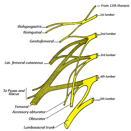
The femoral nerve arises from the Lumbar
Plexus (Ventral rami of L1-L4, the nerve plexus of the lower limb). It arises
from posterior divisions of the ventral rami of the L2-L4 nerve roots, with the
obturator nerve emerging from the anterior divisions of the L2-L4 nerve roots. You
may be wondering why the muscles of the leg have innervation from the lumbar
spinal nerves. Would it not make more sense for them to be innervated by the
sacral nerves as these muscles do not lie in the lumbar region? This can be
explained by the embryological migration of the lumbar musculature down to the
thighs i.e. the quadriceps was originally a lumbar muscle.
The nerve runs through the psoas major, and emerges lateral to the psoas major. It does not supply the psoas major muscle which gets drect supply from the lumbar plexus. It does supply the iliacus muscle. The nerve then runs toward the anterior superior iliac spine, and runs beneath the inguinal ligament before descending in the groin region laterally to the femoral artery and vein. It then divides into a superficial and deep component.
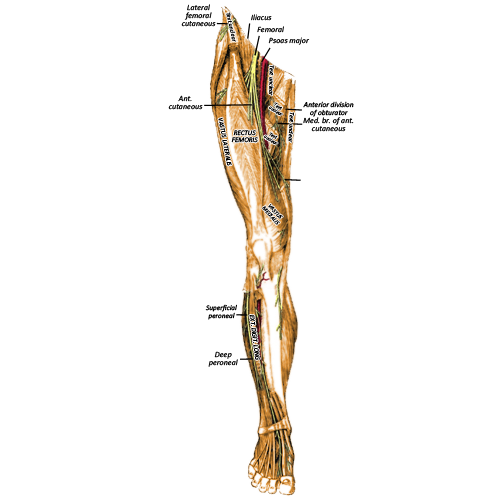
The superficial division supplies sartorius and pectineus and forms the intermediate cutaneous nerve of the thigh, and medial cutaneous nerve of the thigh.
The deep division supplies the anterior compartment of the thigh i.e. quadriceps femoris (vastus medialis, vastus lateralis, vastus intermedius and rectus femoris) and also gives rise to the saphenous nerve. The saphenous nerve runs posterior to the Sartorius muscle in the sub-sartorial (Hunter's) canal, and passes between the sartorius and gracilis muscles. It follows the course of the great saphenous nerve in the medial aspect of the leg and supplies the anteromedial aspects of the knee, leg and foot. The deep division also gives off the infrapatellar branches to the knee, which pierce the fascia lata and sartorius to supply sensation to the skin lying over the patella.
As the femoral nerve supplies the
anterior compartment of the thigh, you may expect it to arise from the anterior
divisions of the ventral rami. It in fact arises from the posterior divisions.
The reason for this can be found in the embryology of the musculature. The
upper limbs remain in their original position, with the flexor compartments on
the anterior surface and the extensor compartments on the posterior surface.
Contrastingly, the lower limb rotate a full 180 degrees, which caused the
flexor compartment to move to the back, and the extensor compartment to move to
the front. The nerve supply to the muscles does not alter, and the artefact of
the original positions of the muscles can be seen in the divisions of the nerve
roots.
Clinical Anatomy
Trauma -
The femoral nerve is at risk from damage following lower limb trauma. The nerve
has a superficial course near the anterior superior iliac spine and can be
crushed under the inguinal ligament. Hip surgery, pelvic surgery, femoral
artery aneurysms, local tumours, haematomas and femoral artery catheterisation
put the nerve at risk. Symptoms include loss of sensation over the medial and
intermediate surface of the thigh, as well as weakened knee extension.
Quick Anatomy
Key Facts
Developmental precursor- Alar and basal plate of L2-L4
Origin-
posterior divisions of anterior rami of L2-L4
Branches-
medial cutaneous nerve of the thigh, intermediate cutaneous nerve of the thigh,
motor branch
Muscles supplied- Quadriceps femoris, pectineus, Sartorius, iliacus.
Dermatome-
medial and intermediate surface of the thigh.
Aide-Memoire
NAVY- this teaches the order of
structures in the femoral neurovasculature from lateral to medial (nerve artery
vein Y-front)
Summary
The
femoral nerve is a branch of the lumbar plexus (the nerve plexus of the lower
lib). It supplies motor innervation to the anterior compartment of the thigh.
It supplies sensation over the anterior and medial thigh and medial surface of
the leg.