Reading Time:
Overview
Embryology provides a basis for understanding anatomy and an explanation of many of the congenital anomalies that are seen in clinical medicine. A very brief overview of the development of the embryo follows.
Once the ovum (egg) has been fertilized by the spermatozoon (sperm), a single cell is formed, called the zygote. This undergoes a rapid succession of mitotic divisions with the formation of smaller cells. The centrally placed cells are called the inner cell mass and ultimately form the tissues of the embryo. The outer cells, called the outer cell mass, form the trophoblast, which plays an important role in the formation of the placenta and the fetal membranes.
The cells that form the embryo become defined in the form of a bilaminar embryonic disc, composed of two germ layers. The upper layer is called the ectoderm and the lower layer, the endoderm. As growth proceeds, the embryonic disc becomes pear shaped, and a narrow streak appears on its dorsal surface formed of ectoderm, called the primitive streak. The further proliferation of the cells of the primitive streak forms a layer of cells that will extend between the ectoderm and the endoderm to form the third germ layer, called the mesoderm.
Ectoderm
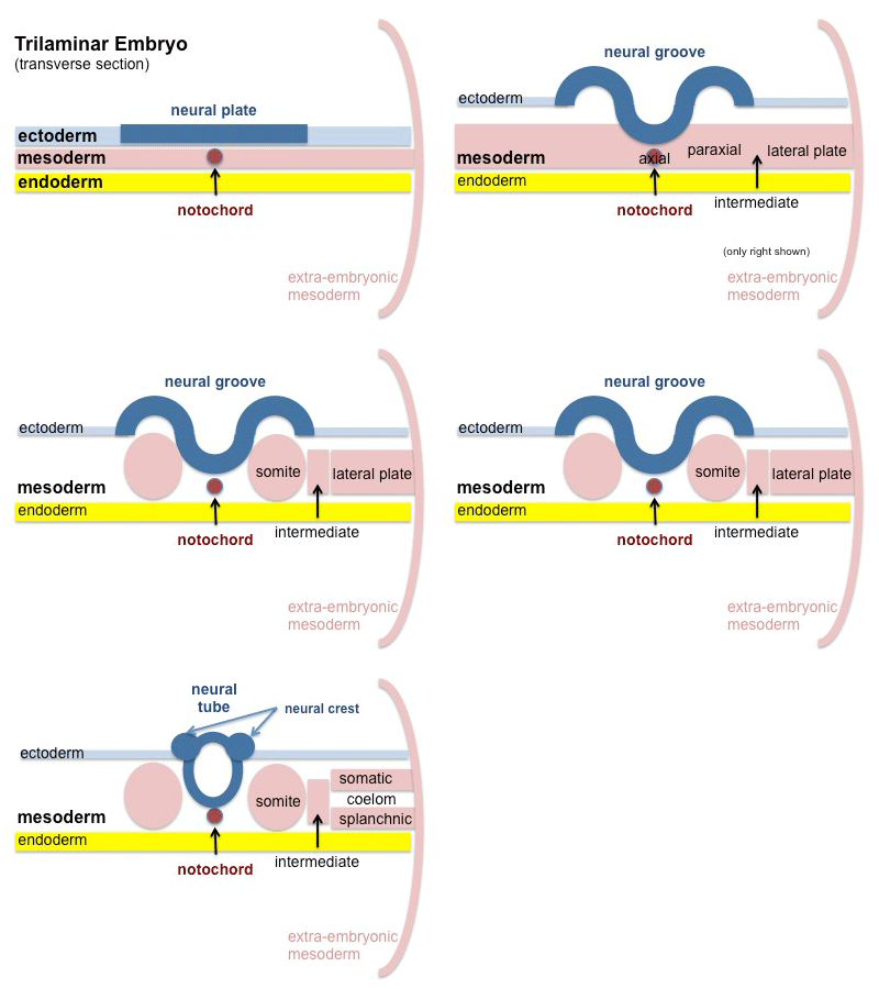
Further thickening of the ectoderm gives rise to a plate of cells on the dorsal surface of the embryo called the neural plate. This plate sinks beneath the surface of the embryo to form the neural tube, which ultimately gives rise to the central nervous system. The remainder of the ectoderm forms the cornea, retina, and lens of the eye and the membranous labyrinth of the inner ear. The ectoderm also forms the epidermis of the skin; the nails and hair; the epithelial cells of the sebaceous, sweat, and mammary glands; the mucous membrane lining the mouth, nasal cavities, and paranasal sinuses; the enamel of the teeth; the pituitary gland and the alveoli and ducts of the parotid salivary glands; the mucous membrane of the lower half of the anal canal; and the terminal parts of the genital tract and the male urinary tract.
Puberty begins between ages 10 and 14 in girls and between 12 and 15 in boys. In the girl at puberty, the breasts enlarge and the pelvis broadens. At the same time, a boy’s penis, testes, and scrotum enlarge; in both sexes, axillary and pubic hair appear.
Racial differences may be seen in the color of the skin, hair, and eyes and in the shape and size of the eyes, nose, and lips. Africans and Scandinavians tend to be tall, as a result of long legs, whereas Asians tend to be short, with short legs. The heads of central Europeans and Asians also tend to be round and broad.
Endoderm
The endoderm eventually gives origin to the following structures: the epithelial lining of the alimentary tract from the mouth cavity down to halfway along the anal canal and the epithelium of the glands that develop from it — namely, the thyroid, parathyroid, thymus, liver, and pancreas—and the epithelial linings of the respiratory tract, pharyngotympanic tube and middle ear, urinary bladder, parts of the female and male urethras, greater vestibular glands, prostate gland, bulbourethral glands, and vagina.
Mesoderm
The mesoderm becomes differentiated into the paraxial, intermediate, and lateral mesoderms. The paraxial mesoderm is situated initially on either side of the midline of the embryo. It becomes segmented and forms the bones, cartilage, and ligaments of the vertebral column and part of the base of the skull. The lateral cells form the skeletal muscles of their own segment, and some of the cells migrate beneath the ectoderm and take part in the formation of the dermis and subcutaneous tissues of the skin.
The intermediate mesoderm is a column of cells on either side of the embryo that is connected medially to the paraxial mesoderm and laterally to the lateral mesoderm. It gives rise to portions of the urogenital system.
The lateral mesoderm splits into a somatic layer and a splanchnic layer associated with the ectoderm and the endoderm, respectively. It encloses a cavity within the embryo called the intraembryonic coelom. The coelom eventually forms the pericardial, pleural, and peritoneal cavities.
The embryonic mesoderm, in addition, gives origin to smooth, voluntary, and cardiac muscles; all forms of connective tissue, including cartilage and bone; blood vessel walls and blood cells; lymph vessel walls and lymphoid tissue; the synovial membranes of joints and bursae; and the suprarenal cortex.
Key Images

Key Info
The key layers are the ecto-, meso- and endoderm. When describing position in the embryo there is a minor change to traditional nomenclatures. Cephalic refers to the head of the embryo, while caudal refers to the tail (inferior) end. The term ventral refers to the anterior (front) aspect of the embryo, while dorsal refers to the posterior (back).
Clinical Anatomy
Birth defects can occur due to a number of clinical reasons. Common defects include palate defects such as cleft palate and cardiac defects such as transposition of the arteries and septal defects.
Neural tube defects include spina bifida and anencephaly.
Summary
The cells that form the embryo become defined in the form of a bilaminar embryonic disc, composed of two germ layers. The upper layer is called the ectoderm and the lower layer, the endoderm. As growth proceeds, the embryonic disc becomes pear shaped, and a narrow streak appears on its dorsal surface formed of ectoderm, called the primitive streak. The further proliferation of the cells of the primitive streak forms a layer of cells that will extend between the ectoderm and the endoderm to form the third germ layer, called the mesoderm.
References
Fitzgerald, M. J. T.: Human Embryology. Harper and Row, Hagerstown, MD, 1978
